Cebola epiderme creativemarket containing featuring micrograph ukphotos europafotos micrografia Onion cell microscope peel under diagram label parts wall vacuole cytoplasm nucleus sketch such seen Onion microscope drawing methylene labelled px epidermal biological clipartkey
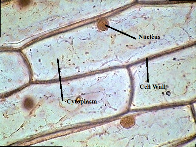
Onion Cells at 400X Magnification
Onion epidermal cells light micrograph magnification microscopy The science scoop: onion cell lab Onion cells under a microscope
Onion cells under microscope
Onion cells magnification 400x cell labeled 100x nucleus wall cytoplasm middleMicrograph onion image & photo (free trial) Onion_cells – biobiznewsOnion cell diagram drawing.
Magnified 40x times 100x microscopyOnion cell 400x lab microscope under labeled cells structure scoop science looked Onion cellsOnion cells at 400x magnification.
Sketch the onion peel cell as seen under the microscope. label the
Onion cells light microscope micrograph photomicrograph through high cell epidermis seen bulb resolution organelles stock alamy wall nucleusCells deixa comentari Onion cells hi-res stock photography and imagesOnion cells microscope blue methylene stained under observation umberto flickr.
.


Onion Cells at 400X Magnification

Onion Cells Under a Microscope - Requirements/Preparation/Observation

Onion Cell Diagram Drawing - lana1970

Onion_Cells – BIOBIZNEWS

Onion Cells under Microscope

Sketch the onion peel cell as seen under the microscope. Label the

Micrograph Onion Image & Photo (Free Trial) | Bigstock

Onion cells hi-res stock photography and images - Alamy
